
Animal study
The overall goal of this project is to investigatemicrocirculation and edema in models of head injury and ischemia.
(1) Experimental head injury
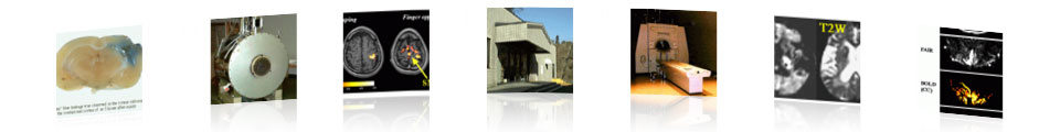
Although it has been considered that edemafollowing cerebral contusion is vasogenic, there are few reportsconcerning diffusion MRI, which is sensitive to discriminate betweencytotoxic and vasogenic edema. We examined the time course ofthe ADC value both in core and surrounding areas of contusionin conjunction with morphological studies by microscopy and electronmicroscopy. In the acute stage, the core of the contusion in thetypical moderate type showed slow diffusion, while the tissuesurrounding the contusion showed fast diffusion. In contrast,the core of the contusion in the severe type showed fast diffusion(green arrow) and tissue the contusion peripheri showed slow diffusion(blue arrow). Light microscopic and electron microscopic examinationsrevealed marked swelling of the axons, dendrites, and glial processesin the areas indicating slow diffusion. The corpus callosum inboth types showed fast diffusion (yellow arrow). Eight days afterinjury, the ADC value of moderate and severe types returned tothe normal level. In conclusion, brain edema in experimental cerebralcontusion consisted of the combination of fast diffusion (vasogenicedema) and slow diffusion (cytotoxic edema) areas.


(Umeda M, et al. Shinkei Gaisho 19: 79-83 [Japanese],1996)
(2) Experimental focal ischemia
(Work in progress)
Last Updated by T.Ebisuon 5/01/98